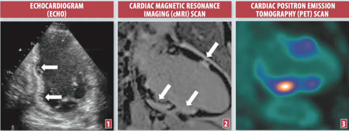
Infiltrative cardiomyopathies are diseases that typically cause the heart muscle to become stiff, dysfunctional and, sometimes, thickened from an accumulation of abnormal material within the muscle itself. This can be fibrosis (scar tissue), inflammatory tissue, a mineral such as iron or an abnormal protein such as amyloid.
To continue reading this article or issue you must be a paid subscriber. Sign in
Subscribe to Heart Advisor
Get the next year of Heart Advisor for just $20. And access all of our online content - over 2,000 articles - free of charge.
Subscribe today and save 38%. It's like getting 5 months FREE!


